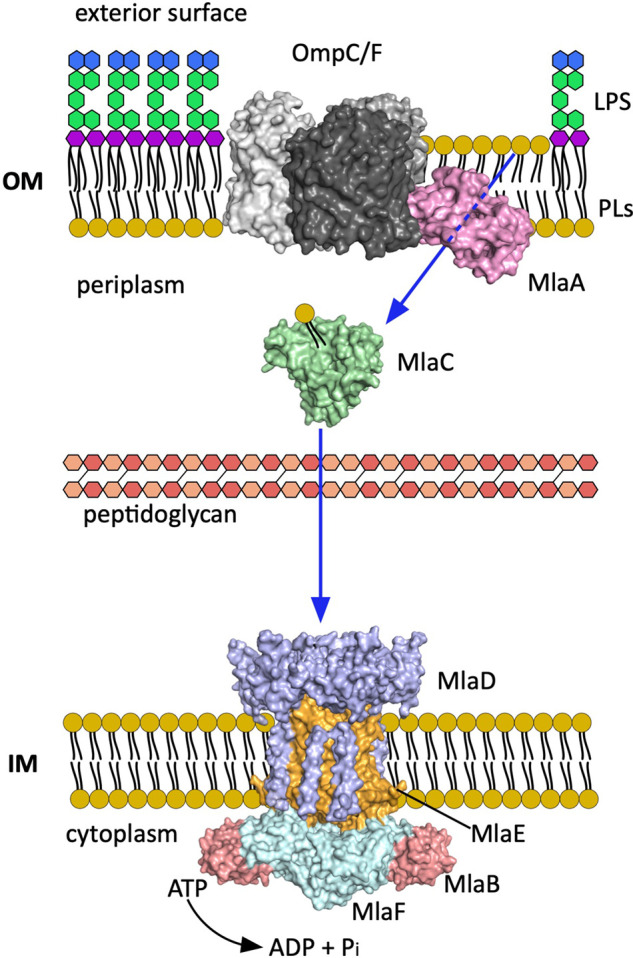
Figure 2. Overview of the Mla pathway.

Localisation of the elements of the Mla system in the cell envelope. Each monomer of the trimeric porin OmpF/C is represented in different shades of grey. The MlaFEDB components are coloured by type of protein and not by monomers. The blue arrows indicate the most accepted PLs movement direction (retrograde). (PDB codes; MlaA–OmpF/C 5NUO [33], MlaC 5UWA [34], MlaFEDB 6XBD [35]).
