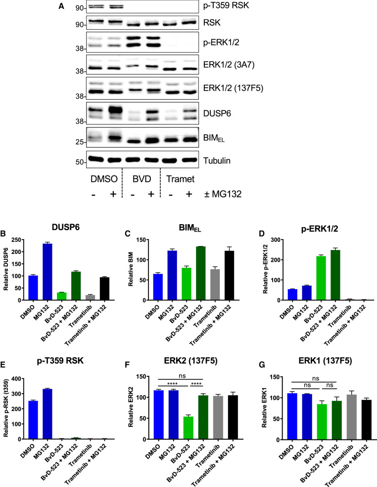
Figure 4. The ERK inhibitor BVD-523 promotes the proteasome-dependent turnover of ERK2.
(A) HCT116 cells were exposed to 0.1% DMSO (control), BVD523 (3 µM) or Trametinib (100 nM) alone or plus (10 µM) MG132 for 24 h. Whole cell lysates were subjected to SDS–PAGE and immunoblotting with the indicated antibodies. Molecular masses in kDa are indicated on the left-hand side. (B–G) Results of Li-Cor quantitative western blotting for (B) DUSP6, (C) BIM, (D) p-ERK1/2 (pThr202/pTyr204-ERK1/2), (E) p-Thr359 p90RSK, (F) ERK2 (with ERK1/2 clone 137F5) or (G) ERK1 (with ERK1/2 clone 137F5). Results are the mean normalised blot quantification ± SEM, n = 3 experiments P-values P < 0.05 **, P < 0.01 *** P < 0.001 **** using one-way analysis of variance with Tukey's multiple comparison test.

