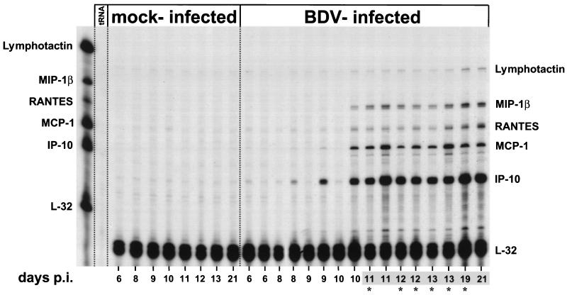
FIG. 1.
Kinetics of expression of various chemokine genes in brains of adult BDV-infected rats. RNA samples (10 μg) from the right brain hemispheres (excluding cerebella) of mock- and BDV-infected rats sacrificed at different times p.i. were subjected to RPA using the rat chemokine probe set. E. coli tRNA (10 μg) was analyzed as a negative control. Positions of the various undigested probes are indicated on the left; positions of the protected probes are given on the right. The autoradiograph was exposed for 2 days. Numbers shaded in gray indicate brains which displayed perivascular infiltrates, as assessed by hematoxylin staining of frozen sections derived from the left brain hemispheres. Asterisks indicate rats which exhibited clinical symptoms of BD.

