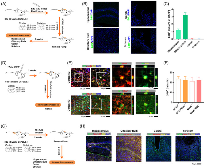
FIGURE 2.

The 5C medium induces direct transdifferentiation around the injury site. (A) Experimental design for intraperitoneal injection of EdU and implantation of osmotic minipump into the brain. (B) Immunofluorescence for EdU (green) in the hippocampus, cortex, olfactory bulb and striatum. (C) Quantification of the percentage of EdU+ cells in DAPI+ cells in the hippocampus, cortex, olfactory bulb and striatum. (D) Experimental design for labelling with GFP‐encoding adenovirus before the 5C medium infusion. (E,F) Immunofluorescence and quantification for the abilities of GFP‐encoding adenovirus to label different types of cells, GFAP+, GFAP+EdU+, NeuN+ and NeuN+EdU+ cells. (G) Experimental design for staining against NeuroD1 and DCX around the injury site 2 weeks after minipump removing. (G) Immunofluorescence for GFAP (green), NeuroD1 (red) and DCX (yellow) in the hippocampus, cortex, olfactory bulb and striatum. Each group included at least three mice and at least four frozen sections from each mouse were analyzed (n ≥ 12). Error bars represented standard errors. Additional statistic information could be found in Table S2.
