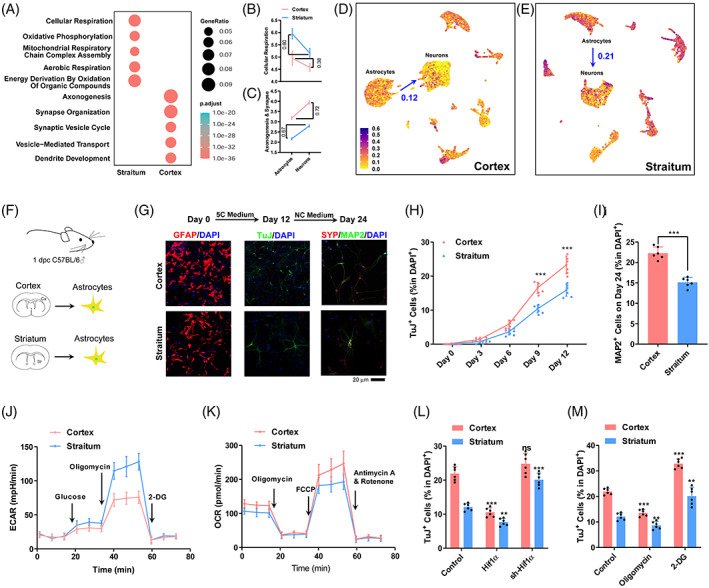
FIGURE 3.

Low glycolysis contributes to the higher transdifferentiation rate in cortex. (A) Gene Ontology studies of differentially expressed genes between cortical and striatal astrocytes. (B,C) Expression changes of genes related to cellular respiration (B) and axonogenesis and synapse (C) during transdifferentiation in the cortex and striatum. (D,E) Expression of genes related to cellular respiration in the cortex (D) and striatum (E) at single cell level. (F) Schematic overview of transdifferentiation in vitro. (G) Representative immunofluorescence images show that astrocytes can be converted into neurons by 5C medium and form mature neurons after incubation with NC medium for additional 12 days. (H,I) The percentage of TuJ+ cells and MAP2+ cells in DAPI+ cells. (J,K) Energy metabolism of primary astrocytes was analysed with a Seahorse instrument. (L,M) Efficiency of astrocyte transdifferentiation in the cortex and striatum after OXPHOS changes controlled by Hif1α gene up/downregulation and drugs (2‐DG/oligomycin). Experiments were repeated for at least six times (n ≥ 6) except scRNA‐seq from previous report. Error bars represented standard deviations. One‐way ANOVA and two‐way ANOVA were used for L,M and H, respectively, Student two‐tailed t‐test was used for I. Additional statistic information could be found in Table S2.
