
FIGURE 1.
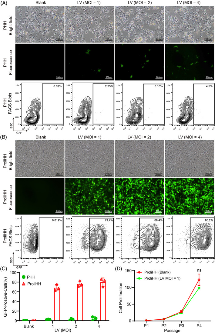
Proliferating human hepatocytes (ProliHHs) are highly susceptible to lentiviral infection. (A) Representative images of primary human hepatocytes (Lot: MRW) in brightfield and fluorescence microscope. Hepatocytes were infected with Empty‐lentivirus (LV) expressing green fluorescent protein (GFP) at multiplicities of infections (MOIs) of 0, 1, 2 and 4 for 3 days in Dulbecco's Modified Eagle Medium. Scale bars, 200 μm. Flow cytometry was performed 48 h after LV transduction. (B) Representative images of ProliHHs (lot: JFC) in brightfield and fluorescence microscope. ProliHHs were infected with Empty‐LV expressing GFP at MOIs of 0, 1, 2 and 4 for 3 days in hepatocyte medium (HM). Scale bars, 200 μm. Flow cytometry was performed 48 h after LV transduction. (C) Percentage of GFP‐positive cells under different titers of Empty‐LV expressing GFP were analysed by the flow cytometry. Data are means ± SD (n = 3 replicates). (D) Growth curves of Empty‐LV‐infected ProliHHs (MOI = 1) were analysed at indicated passages in HM. ProliHHs without LV infection were used as a control. The data are shown as the mean ± SD. NS > 0.05, Student's t test.