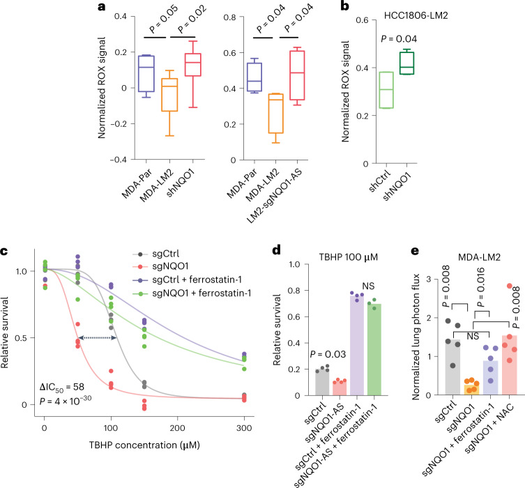
Fig. 6. NQO1 protects cancer cells from oxidative stress and ferroptosis.
a, ROS measured using the CellROX assay in MDA-Par, MDA-LM2, MDA-LM2 NQO1 knockdown and MDA-LM2 NQO1-AS knockdown cells. n = 4 independent cell cultures. b, ROS measured using the CellROX assay in HCC1806-LM2 NQO1 knockdown and control cells. n = 4 independent cell cultures. c, TBHP dose–response in MDA-LM2 NQO1 knockdown and control cells, with or without pretreatment with ferrostatin-1. n = 3 independently treated cell cultures. d, Relative survival of MDA-LM2 NQO1-AS knockdown and control cells after TBHP treatment, with and without pretreatment with ferrostatin-1. n = 4 independently treated cell cultures. e, In vivo lung colonization by MDA-LM2 control and NQO1 knockdown cells, with and without pretreatment with ferrostatin-1 or NAC, as measured by luciferase assay. n = 5 mice per cohort. In a,b the boxplots represent the median values and quartiles, and the whiskers represent the maximum and minimum values. The P values in a,b were calculated using an unpaired, one-tailed t-test. The P value in c was calculated using the drc package in R. In d,e P values were calculated using a one-tailed Mann–Whitney U-test.

