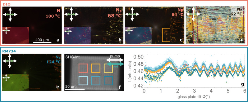
Fig. 1. Photopatterned millimeter range uniform polar domains.
a–c 1.3 × 0.7 mm photopatterned uniform domains in DIO in the N phase (a), in the NF phase before final structural relaxation (b), and in the NF phase after structural relaxation in a 3 µm thick cell (c). The main images show the patterned aligning direction of the investigated area (horizontal as indicated by bidirectional green arrow) aligned with the crossed polarizers (white arrows), while the inset shows the same area under 20° anticlockwise rotation. Microphotographs show a uniform structure with some defects (see Fig. 2) arising from the unpatterned edges of the structured area. d Same photopatterned structure in a region of a cell where one of the surfaces has interdigitated electrodes shows that the absence of conductive surfaces leads to an uncontrolled division in ferroelectric domains due to uncompensated charges and polarization distortions due to local impurities. Dashed maroon lines in d mark the position of the electrodes. In the broad region between them, only one of the confining cell surfaces has an ITO electrode. e Observations of the same photopatterned structure filled with RM734. f SHG-I image of the RM734 uniform structure and g SHG interferogram corresponding to the highlighted areas in (f). Measurements in the different areas show the same phase, indicating that the polarization direction is the same throughout the uniform pattern. Error bars smaller than the point size.

