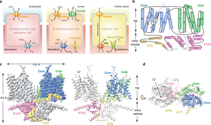
Fig. 1. Cellular functions and cryo-EM structure of Pendrin.
a Cellular functions of pendrin. In the inner ear, pendrin mediates the exchange of HCO3− and Cl−; in β-intercalated cells, it participates in urinary HCO3− excretion and tubular Cl− reabsorption; and in the thyroid, pendrin is involved in apical I− transport. NIS, sodium iodide symporter. b Topology of Pendrin. The N-terminal (NTD), core region, gate region, and STAS domains are colored in yellow, blue, green, and pink, respectively. The anion-binding pocket is marked by a dashed circle in cyan. The key residues around the pocket are labeled. c Cryo-EM map and structural model of mouse pendrin-Cl. One protomer is colored in grey, and the other is colored as (b). d View of c from outside the membrane. The Cl− are shown as spheres in magenta.

