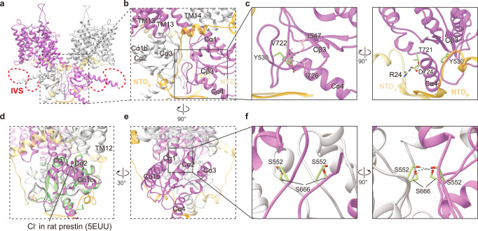
Fig. 4. STAS domain of pendrin.
a Structure model of pendrin-Cl. Protomer A is colored in orchid and orange, protomer B is colored in light grey and light yellow. b The dimerization interface around the STAS domain. c Details of STAS domain. Y530, T721, and D724 and their interactions are indicated by dot lines. d Comparison of STAS domain of pendrin-Cl with that of rat prestin (crystal structure). Rat prestin-Cl crystal structure (PDB 5EUU) is colored light green. Cl− in the hypothetic pre-binding site is pointed out. e A 90° rotated view of (b). f Details of the interface between two STAS domains. Hydrogen bonds between S552 and S666 are indicated.

