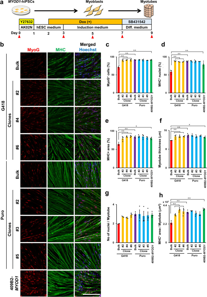
Figure 2.
Bulk MYOD1-hiPSCs established with puromycin selection achieved efficient myogenic differentiation and myotube maturation similar to that achieved by clonally established MYOD1-hiPSCs. (a) Schematic presentation of skeletal muscle differentiation from MYOD1-hiPSCs. Samples were collected at the times indicated with the closed triangles. (b) ICC analysis of myotubes derived from bulk and clonal MYOD1-hiPSCs established with G418 or puromycin selection and control 409B2-MYOD1-hiPSCs for the expression of MyoG and MHC at day 9 of differentiation. The nuclei were stained with Hoechst 33258. Scale bar, 200 μm. (c–e) Quantitative analysis of the parameters for skeletal muscle differentiation, including the proportions of MyoG+ cells among total cells (c), the proportions of MHC+ nuclei among total nuclei (d), and the proportion of MHC+ area in the total area (e). Puro-bulk MYOD1-hiPSCs exhibited differentiation potential similar to that of clonal MYOD1-hiPSCs and control 409B2-MYOD1-hiPSCs, whereas G418-bulk MYOD1-hiPSCs exhibited lower differentiation potential than the other MYOD1-hiPSCs. (f–h) Quantitative analysis of the parameters for myotube maturation, including the myotube thickness (f), number of nuclei per myotube (g), and MHC+ area per myotube (h). Puro-bulk MYOD1-hiPSCs exhibited myotube maturation potential similar to that of clonal MYOD1-hiPSCs and control 409B2-MYOD1-hiPSCs, whereas G418-bulk MYOD1-hiPSCs exhibited lower myotube maturation potential than the other MYOD1-hiPSCs. The data are presented as the mean ± SEM, n = 3. *p < 0.05, **p < 0.01. ANOVA followed by post hoc Bonferroni test.

