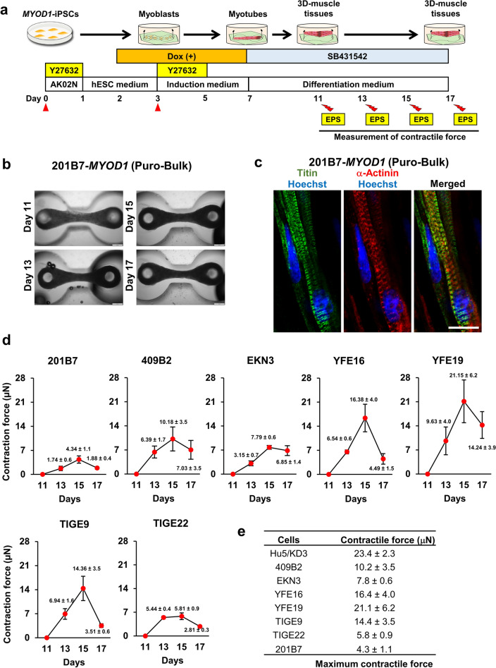
Figure 7.
3D muscle tissues fabricated from Puro-bulk MYOD1-hiPSCs exhibited contractile force, indicating the functionality of the hiPSC-derived muscle tissues. (a) Schematic of the fabrication of 3D muscle tissues. On day 3 of differentiation, differentiating cells were dissociated and replated on microdevices. On day 11, muscle tissues were pulled up at the top of the pillar and processed for measurement of contractile force elicited by EPS at days 11, 13, 15 and 17. (b) Brightfield top-view images of muscle tissues derived from Puro-bulk 201B7-MYOD1-hiPSCs at days 11, 13, 15, and 17. Scale bar, 500 μm. (c) IHC analysis of fabricated muscle tissues from Puro-bulk 201B7-MYOD1-hiPSCs for Titin and α-Actinin at day 17 of differentiation. Sarcomere formation was clearly observed in Puro-bulk 201B7-MYOD1-hiPSC-derived muscle tissues. The nuclei were stained with Hoechst 33258. Scale bar, 20 µm. (d) Measurement of the contractile force of the muscle tissues derived from seven hiPSC clones (201B7, 409B2, EKN3, YFE16, YFE19, TIGE9, and TIGE22) at days 11, 13, 15, and 17 of differentiation. The maximum contractile force is recorded at day 15. The data are presented as the mean ± SEM, n = 4. (e) Comparison of the maximum contractile force of muscle tissues derived from seven hiPSC clones (201B7, 409B2, EKN3, YFE16, YFE19, TIGE9, and TIGE22) and Hu5/KD3 cells.

