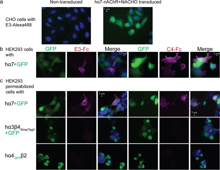
Fig. 3.
VHH characterization by immunofluorescence. Dapi, shown in blue, stains the cells’ nucleus. Alexa Fluor™ 647, a red wavelength, is colored as magenta. Images are representative of at least n = 4. a Merged images (Dapi + GFP) of CHO cells (left) and with stable expression of hα7-nAChR (right); immunostained with 1 µg/mL of conjugated E3-CSA-Alexa Fluor™ 488. b Images (GFP, Alexa Fluor™ 647, Merged [GFP + Alexa Fluor™ 647 + Dapi]) of non-permeabilized HEK 293 cells transfected with hα7-nAChR immunostained with E3-Fc (left) and C4-Fc (right), demonstrating an extracellular binding. Cytoplasmic GFP indicates efficiently transfected cells. c Images (GFP, Alexa Fluor™ 647, Merged [GFP + Alexa Fluor™ 647 + Dapi]) of permeabilized HEK 293 cells expressing hα7-, hα3β4StrepII- and hα4GFPβ2-nAChRs immunostained using E3-Fc (left) and C4-Fc (right). For hα7 and hα3β4Strep, cytoplasmic GFP indicates efficiently transfected cells. The nanobodies were detected by an anti-human IgG coupled to Alexa Fluor™ 647 (red). Identical exposure times were used to visualize each channel on all conditions

