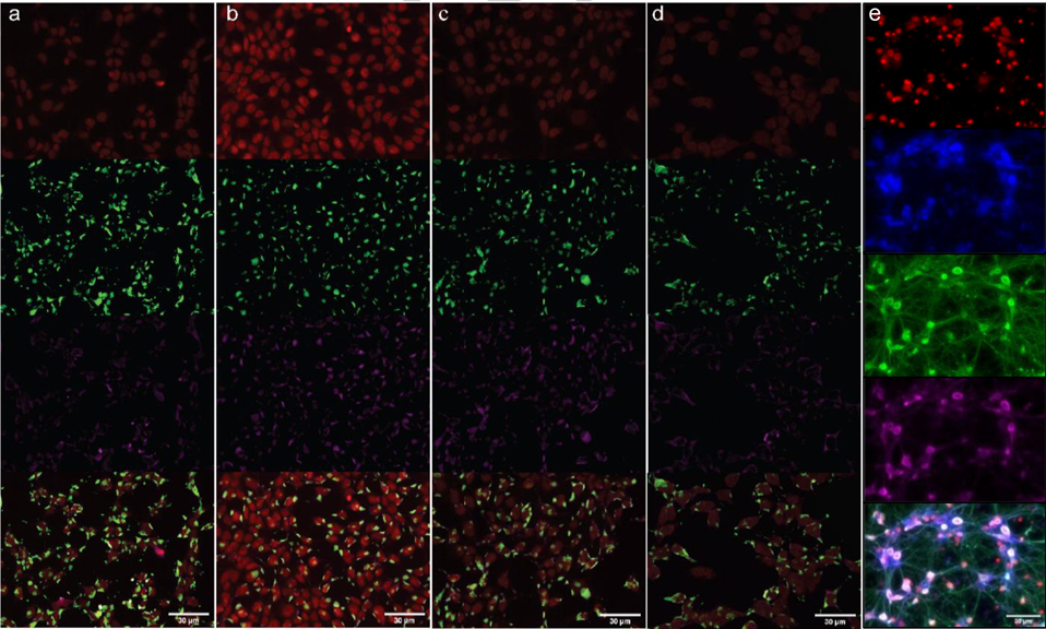
Figure 5.
Immunofluorescence staining of HEK293T cells treated with (a) 10 μM puromycin (1), (b) 10 μM thpuromycin (2), (c) 10 μM AzThpuromycin (3), and (d) 10 μM F2AzThpuromycin (4) for 2 hours. Image channels top to bottom: DRAQ5 (nuclear stain), anti-puromycin (primary) and GAMAF488 (secondary), HPO-0100 (anti-ribosome, primary) and DAH Cy3 (secondary), overlay. (e) Fixed primary rat hippocampal neuron images stained with DRAQ5 (top), compound 4 (second row), anti-puromycin (third row), and anti-ribosome (fourth row). Images are overlayed (bottom).

