Figure 3.

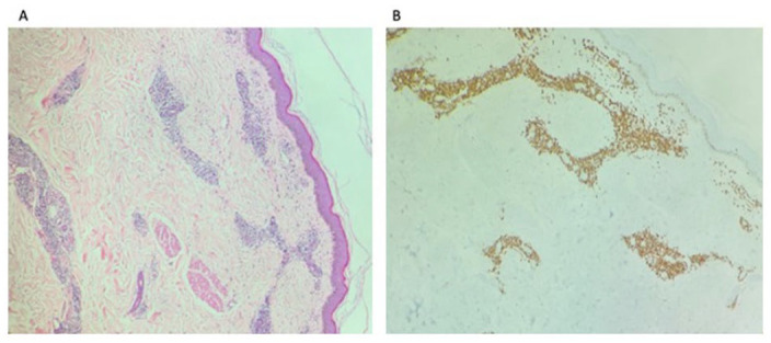
Atypical perivascular T-cell infiltrates (A) and were positive for CD3, CD4, CD5, and CD7 and negative for CD8, CD30, and CD56 (B) consistent with cutaneous involvements by the diagnosis of T-prolymphocytic leukemia (T-PLL).

Atypical perivascular T-cell infiltrates (A) and were positive for CD3, CD4, CD5, and CD7 and negative for CD8, CD30, and CD56 (B) consistent with cutaneous involvements by the diagnosis of T-prolymphocytic leukemia (T-PLL).