Fig. 5.

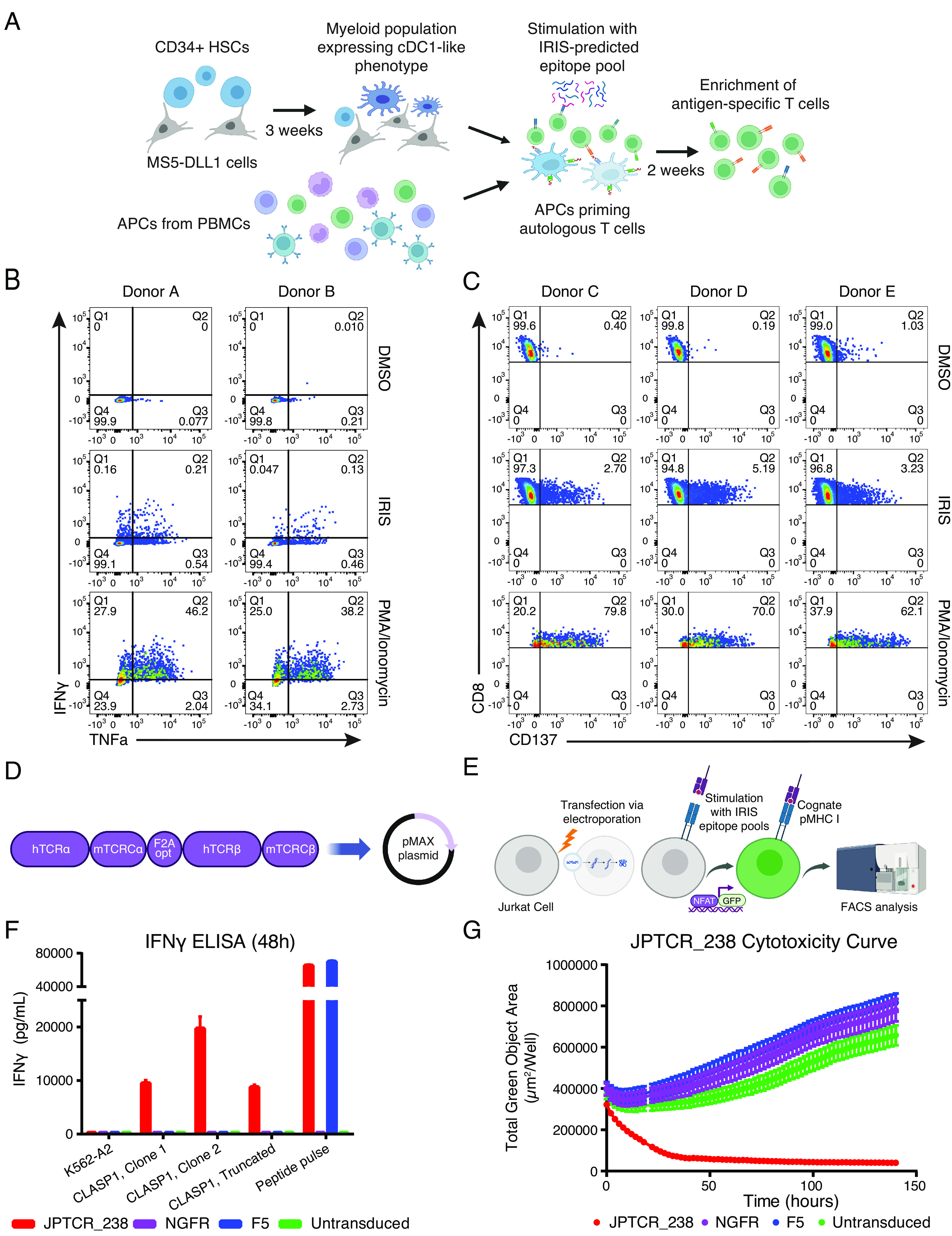
Isolation and characterization of TCRs reactive to IRIS-predicted NEPC epitopes. (A) IRIS-epitope priming using two APC systems: (1) conventional type 1 dendritic cell (cDC1)-like cells differentiated from autologous CD34+ hematopoietic stem cells (HSCs), and (2) existing APCs from PBMCs. (B) Example of reactive T cell populations primed with a DMSO negative control, IRIS epitope pool, or PMA/Ionomycin using the CLInt-seq TNFα/IFNγ intracellular marker staining strategy. (C) Example of reactive T cell populations primed with a DMSO negative control, IRIS epitope pool, or PMA/Ionomycin by the CD137 surface marker staining strategy. (D) Overview of the cloning strategy for TCRα/β chains in the pMAX system for Jurkat-NFAT-GFP screening. (E) Overview of the Jurkat-NFAT-GFP reporter system. (F) IFNγ ELISA of one specific TCR (JPTCR_238) targeting an IRIS-predicted AS-derived epitope in CLASP1, when co-cultured with K562-A2-GFP single-cell clones transduced to express a full-length or truncated CLASP1 protein isoform. Error bars indicate SD (n = 3). JPTCR_238: an isolated TCR targeting an IRIS-predicted epitope in CLASP1; F5: a clinically tested TCR targeting the MART1 melanoma antigen; NGFR: empty vector with no introduced TCR as a negative control; Untransduced: untransduced as a negative control. (G) Cytotoxicity analysis by live cell imaging of a K562-A2-GFP single-cell clone transduced with a full-length CLASP1 protein isoform containing an IRIS-predicted epitope targeted by JPTCR_238. F5 TCR, NGFR (no TCR introduced), and untransduced were used as negative controls.
