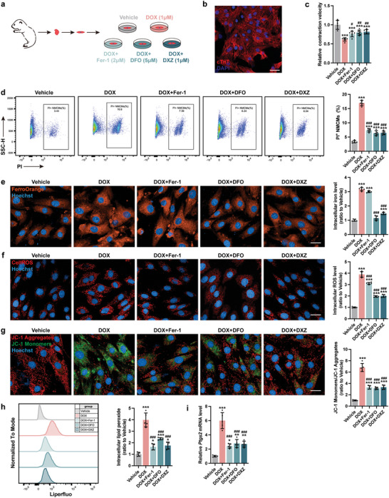
Figure 2.

Blocking myocardial ferroptosis significantly alleviates DoIC in vitro. a) Experiment design for DoIC in neonatal murine cardiomyocytes (NMCMs) and Fer‐1, DFO, and DXZ were used for anti‐ferroptosis treatment prior to DOX challenge. b) Representative micrographs of isolated neonatal murine cardiomyocytes (NMCMs) stained with cTnT (scale bars, 50 µm). c) Contractile velocity of NMCMs extrapolated from live cell imaging (n = 6). d) The percentage of PI+ NMCMs were calculated using flow cytometry (n = 5). e) Representative micrographs and quantification of intracellular iron level (FerroOrange staining) in NMCMs (scale bars, 20 µm; n = 5). f) Representative micrographs and quantification of intracellular ROS (CellROS staining) in NMCMs (scale bars, 20 µm; n = 5). g) Representative micrographs and quantification of mitochondrial membrane potential (JC‐1 staining) in NMCMs (scale bars, 20 µm; n = 5). h) Quantification of intracellular lipid peroxide in NMCMs by flow cytometry (n = 5). i) Ptgs2 mRNA expression in NMCMs (n = 5). The data are expressed as mean ± SD and analyzed using one‐way ANOVA followed by Tukey's post hoc test, *p < 0.05, **p < 0.01, and ***p < 0.001 versus Vehicle group; #p < 0.05, ##p < 0.01, and ###p < 0.001 versus DOX group.
