Figure 6.
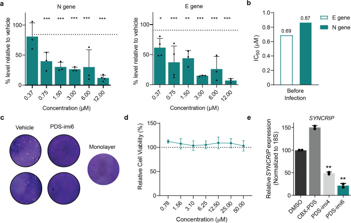
Antiviral effects of PDS-imi6. (a) The percentage inhibition of viral replication normalized to vehicle treated cells (dashed line) after incubation with increasing concentrations of PDS-imi6. Viral replication was assessed 24 h after infection [multiplicity of infection (MOI) of 0.05] based on E and N genes (n = 3). Mean ± SD of triplicates is shown, and differences between means with p < 0.01 are indicated. (b) Calculated IC50 values of PDS-imi6 were determined by quantifying E or N gene. (c) 24-h treatment with PDS-imi6 at 6 μM showed a decreased number of viral plaques in comparison to vehicle control. Images are representative of 4 independent experiments. (d) PDS-imi6 did not show cytotoxicity in VERO-CCL-81 cells (n = 3). (e) RT-qPCR validation of degradation specificity in a cellular system using MOLM-13 cells treated with 5 μM of CBX-PDS, PDS-imi4, and PDS-imi6 or vehicle for 24 h (n = 3). *p < 0.05; **p < 0.01; ***p < 0.001; two-tailed paired t tests versus control.

