Figure 10.
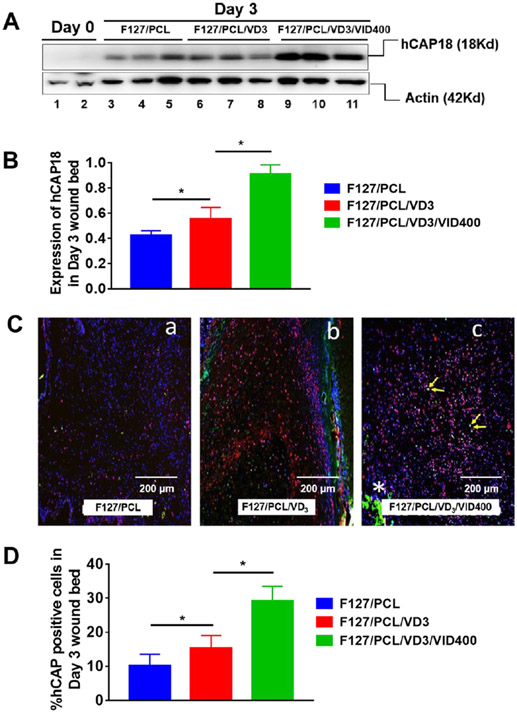
1,25(OH)2D3 and 1,25(OH)2D3/VID400 nanofiber membranes promote LL-37 expression in the CAMPTg/Tg:KO/KO transgenic mouse wound model. (A) Induction of hCAP18 expression in Day 3 skin wounds post treatment with nanofibers containing 1,25(OH)2D3 and 1,25(OH)2D3/VID400. Protein samples were extracted from day 3 skin wounds of CAMPTg/Tg:KO/KO mice treated with F127/PCL only, F127/PCL/VD3 and F127/PCL/VD3/VID400. Western blots were performed using specific anti-hCAP18 antibody. Actin was used as a loading control. (B) Induction of hCAP18 expression in day 3 skin wounds post treatment with nanofibers containing 1,25(OH)2D3 and 1,25(OH)2D3/VID400. Significant induction of hCAP18 was observed in 1,25(OH)2D3 and 1,25(OH)2D3/VID400 loaded nanofibers compared to the PCL control. (C) Immunofluorescence staining of hCAP18/LL-37 protein (in red) and F4/80 (green) on day 3 samples post wounding in the presence of F127/PCL fibers alone and with 1,25(OH)2D3, as well as 1,25(OH)2D3/VID400. Nuclei were counterstained with DAPI (in blue). (a) F127/PCL; (b) F127/PCL/VD3 and (c) F127/PCL/VD3/VID400. Increased number of hCAP18+ cells were detected in the wound bed of F127/PCL/VD3 treated skin wounds, which was further increased in the skin wounds with F127/PCL/VD3/VID400 nanofibers. (D) Quantitative analysis of the number of hCAP18+ cells detected in the wound bed in day 3 for skin wounds with post-treatment of nanofibers containing 1,25(OH)2D3 and 1,25(OH)2D3/VID400. F127/PCL: PCL/pluronic F-127 blend nanofibers. F127/PCL/VD3: 1,25(OH)2D3 loaded PCL/pluronic F-127 blend nanofibers. F127/PCL/VD3/VID400: 1,25(OH)2D3 and VID400 coloaded PCL/pluronic F-127 blend nanofibers. (*p<0.05)

