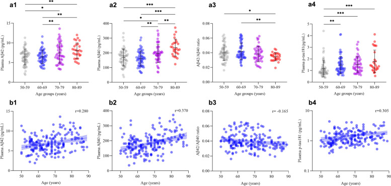
Fig. 1.
Comparison of plasma levels of amyloid-β 42, amyloid-β 40, amyloid-β 42 to amyloid-β 40 ratio, and phosphorylated tau181 among age groups. a (1–4) Interleaved scatter plots show plasma amyloid-β 42, amyloid-β 40, amyloid-β 42 to amyloid-β 40 ratio, and phosphorylated tau181 levels in different age groups. b (1–4) Linear regression plots show the correlations between age and plasma amyloid-β 42, amyloid-β 40, amyloid-β 42 to amyloid-β 40 ratio, and phosphorylated tau181 levels. *P ≤ 0.05, **P ≤ 0.01, ***P ≤ 0.001. Abbreviations: Aβ, amyloid-beta protein; t-tau, total tau; p-tau181, tau phosphorylated at threonine 181

