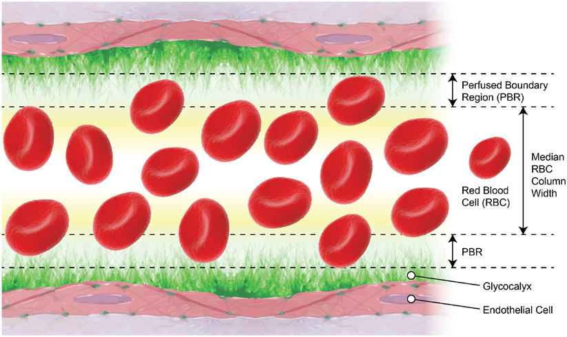
Figure 7.
Schematic representation of the perfused boundary region (PBR) that allows for assessment of in vivo endothelial glycocalyx thickness. Flowing red blood cells (RBC) form a column as they travel within the vascular lumen. Blood velocity and the electrostatic charges of the endothelial glycocalyx as well as those of RBCs form a PBR, where only a few RBCs occasionally penetrate. The size of the median RBC column width subtracted from the vascular luminal diameter represents a measure of the PBR and the thickness of the endothelial glycocalyx. The figure was modified from (56).

