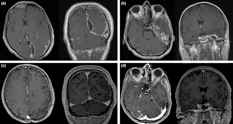
Figure 3.

(a) First MRI of the patient when he became symptomatic showing interhemispheric SDE. (b) Initial MRI of the patient showing temporal SDE. (c and d) Follow-up MRI of the patient 2 months after discharge, without residual empyema can be seen both in interhemispheric space and temporal lobe.
