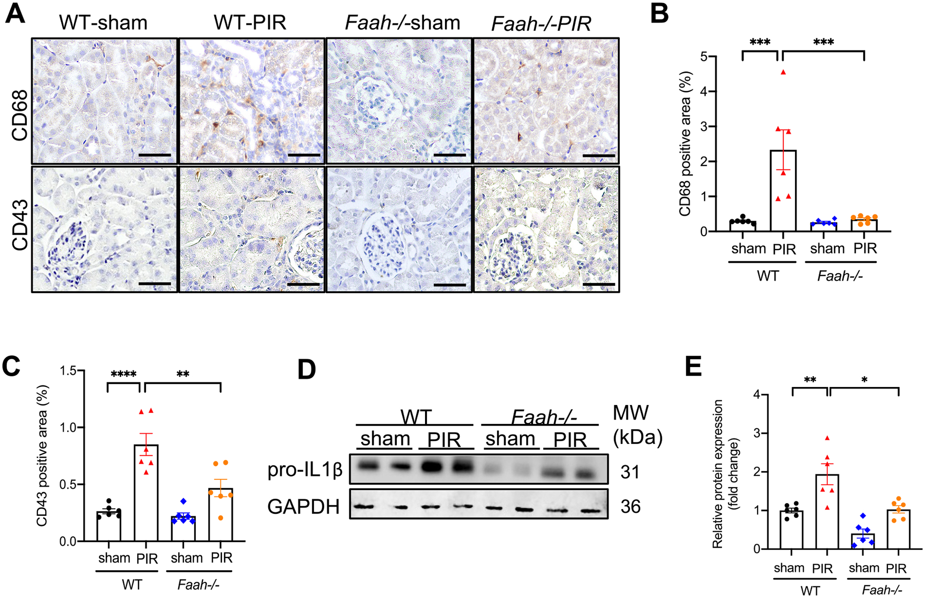
Fig. 4.

Effect of FAAH KO on PIR-induced inflammatory responses in kidneys. (A) Representative immunohistochemical staining showing CD68 (a macrophage marker) and CD43 (a T-cell marker) in kidney tissues. (B) Percentage of positive area of CD68 and (C) CD43. At least 10 fields for each sample were evaluated for all results. Black bar = 100 μm. (D-E) Representative immunoblots and quantitative data (n = 6) showing the level of proinflammatory marker, pro-IL1β in WT and Faah−/− mice. All values were normalized to WT-sham. *P < 0.05, **P < 0.01, *** P < 0.001, **** P < 0.0001 by two-way ANOVA Tukey’s multiple comparisons test.
