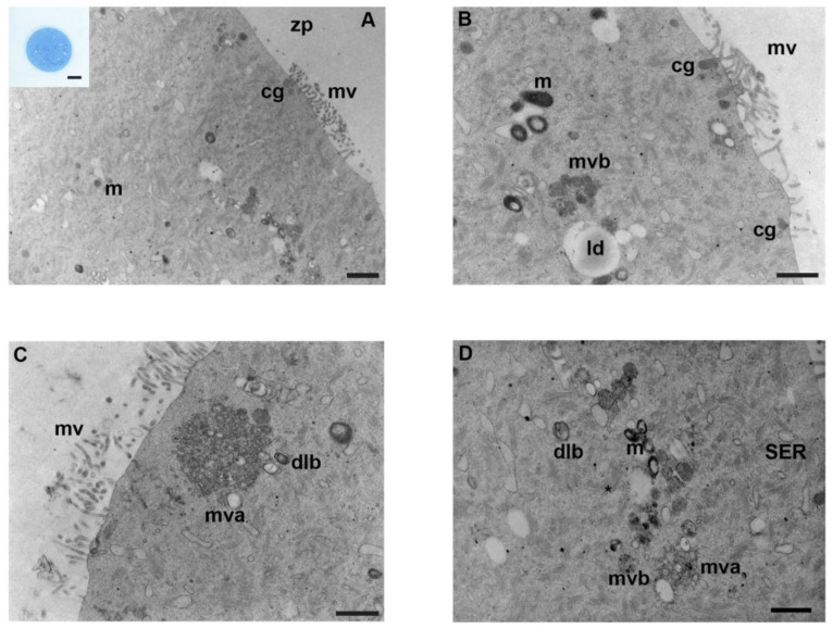
Figure 1.
Ultrastructure of mouse oocytes in the controls group. Representative TEM micrographs showing in (A) the general morphology of the cortical region in MII mouse oocytes, microtopography of intracellular organelles, and microvillar processes. Round/ovoid mitochondria (m) and cortical granules (cg) are visible; zp: zona pellucida; mv: microvilli (TEM, bar: 1 µm). Inset in (A): a representative image of a semithin section of mouse oocyte (LM, Mag: 40×). (B) High magnification, cortex of mouse oocytes evidence clusters of mitochondria (m), lipid droplets (ld), multivesicular bodies (mvb), cortical granules (cg), and regular distribution of microvilli (mv) on the oolemma (TEM, bar: 800 nm). (C) Multivesicular aggregates (mva) are visible in the cortex, with dense lamellar bodies (dlb), at high magnification. Notice long and thin microvilli (mv) (TEM, bar: 800 nm). (D) Portion of ooplasm showing cell organelles: mitochondria (m) with electron-dense cristae, accompanied by multivesicular bodies (mvb), multivesicular aggregates (mva). Dense lamellar bodies (dlb), SER and the extensive fibrillar matrix of cytoplasmic lattice (*) are observed (TEM, bar: 600 nm).

