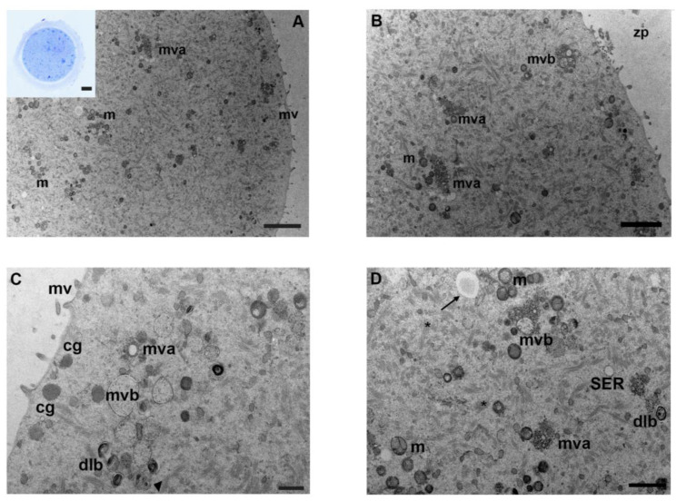
Figure 2.
Ultrastructure of mouse oocytes in mancozeb 0.001 µg/mL group. (A) Low magnification of TEM micrographs from MII mouse oocytes showing high preservation of cell organelles, homogeneously distributed in the cytoplasm. Clustered mitochondria (m) and numerous multivesicular aggregates (mva) are visible; mv: microvilli.(TEM, bar: 2 µm). Inset in (A): a representative semithin section of mouse oocyte (LM, Mag: 40×). (B) Representative TEM image of the cortical region in MII mouse oocytes. Clusters of mitochondria (m), with electron-dense cristae and matrix, are visible, accompanied by multivesicular aggregates (mva) and multivesicular bodies (mvb). zp: zona pellucida (TEM, bar: 500 nm). (C) At high magnification, a small portion of the cortex evidences multivesicular bodies (mvb), with dense lamellar bodies (dlb) and multivesicular aggregates (mva). Cortical granules (cg) are linearly arranged below the oolemma. Note multivesicular bodies (mvb) and dense lamellar bodies (dlb) are in close association with an autophagic-like vesicle (arrowhead). Short and thick microvilli (mv) are observed (TEM, bar: 500 nm). (D) Micrographs of cytoplasmic ultrastructure in mouse oocyte. Image shows different cytoplasmic structures as mitochondria (m), with a round or oval shape and visible double membranes, multivesicular bodies (mvb) and aggregates (mva), dense lamellar bodies (dlb), and fibrillar matrix of cytoplasmic lattice (*). Immature autophagic-like vesicle delimited by a double membrane and a wider lumen (arrow). SER, with small vesicles, is also visible (TEM, bar: 1 µm).

