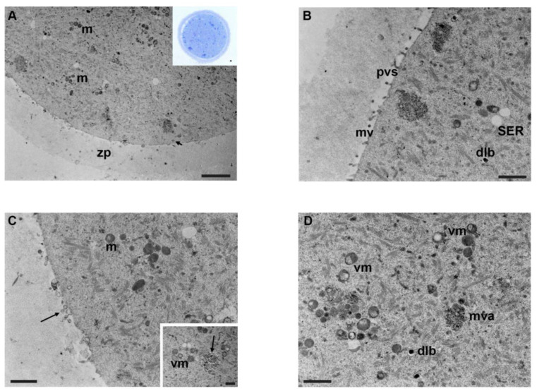
Figure 4.
Ultrastructure of mouse oocytes in mancozeb 0.1 µg/mL group. (A) TEM micrograph showing general morphology of MII oocyte. Few organelles are visible in the cytoplasm; m: mitochondria; zp: zona pellucida. Rare cortical granules are visible (arrow) (TEM, bar: 2 µm). Inset in (A): semithin section of mouse oocytes (LM, Mag: 40×). (B) A portion of the cortical region showing nonhomogeneous and short microvilli (mv) protruded in the perivitelline space (pvs), SER, and dense lamellar bodies (dlb) (TEM, bar: 1 µm). (C) Cortex of mouse oocytes with few organelles, sporadic clusters of mitochondria (m). Notice the presence of extracellular materials and debris (arrow) in the perivitelline space. (TEM, bar: 1 µm). Inset in (C): Mature autophagic-like vesicles (arrow) enclosed in a single membrane, containing material of unrecognizable origin and vacuolated mitochondria (vm) (TEM, bar 600 nm). (D) TEM image of ooplasm showing vacuolated mitochondria (vm), dense lamellar bodies (dlb), multivesicular aggregates (mva), and abundant cytoplasmic lattice (*) (TEM, bar: 1 µm).

