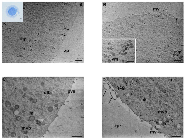
Figure 5.
Ultrastructure of mouse oocytes in mancozeb 1 µg/mL group. (A) Representative TEM micrograph of the cortical region showing low cell organelles density. Few mitochondria (m) are visible; cortical granules are absent. Notice the lack of microvilli (arrow) on the oolemma; zp: zona pellucida (TEM, bar: 2 µm). Inset in (A): semithin section of mouse oocytes, with condensed organelles in one pole (LM, Mag: 40×). (B) A portion of the cortical region with a non-homogeneous distribution of organelles beneath the oolemma and short and thick microvilli (mv). Exocytotic vesicles (arrow) are visible in the perivitelline space; mitochondria (m) (TEM, bar: 1 µm). Inset in (B): groups of mitochondria (m) and vacuolated mitochondria (vm) (TEM, bar: 1 µm). (C) High magnification of cortex part with more organelles present; mitochondria (m) with evident cristae, dense lamellar bodies (dlb), and multivesicular bodies (mvb); pvs: perivitelline space; (*): cytoplasmic lattice; (TEM, bar: 1 µm). (D) Representative TEM micrographs of the cortical region in mouse oocyte. Multivesicular bodies (mvb) are visible. Few cortical granules (cg) are present. Microvilli (mv) are short, and irregularly distributed on the oolemma. Extracellular material, exosomes, and debris (arrow) are detected in the perivitelline space; zp: zona pellucida; (TEM, bar: 1 µm).

