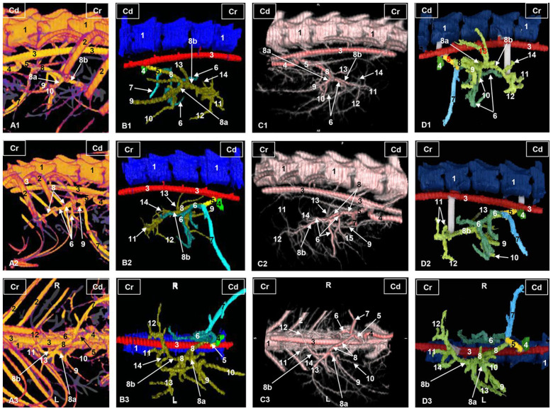
Figure 2.
Three-dimensional reconstructions images of the liver arterial system in cats. (A) Amira volume rendering. (B) Amira segmentation reconstruction. (C) OsiriX volume rendering. (D) 3D printing. (A1–D1) Right side view. (A2–D2) Left side view. (A3–D3) Ventral view. R: right side; L: left side; Cr: cranial; Cd: caudal; 1: thoracic vertebrae; 2: ribs; 3: thoracic aorta—descending aorta; 4: cranial mesenteric artery; 5: celiac artery; 6: left gastric artery; 7: splenic artery; 8: hepatic artery; 8a: hepatic artery, right branch; 8b: hepatic artery, left branch; 9: hepatic artery, branch for the right lateral lobe; 10: hepatic artery, branch for the right medial lobe; 11: hepatic artery, branch for the left lateral hepatic lobe; 12: hepatic artery, branch for the left medial hepatic lobe and quadrate hepatic lobe; 13: hepatic artery, branch for the caudate hepatic lobe (caudate process); 14: hepatic artery, branch for the caudate hepatic lobe (papillary process).

