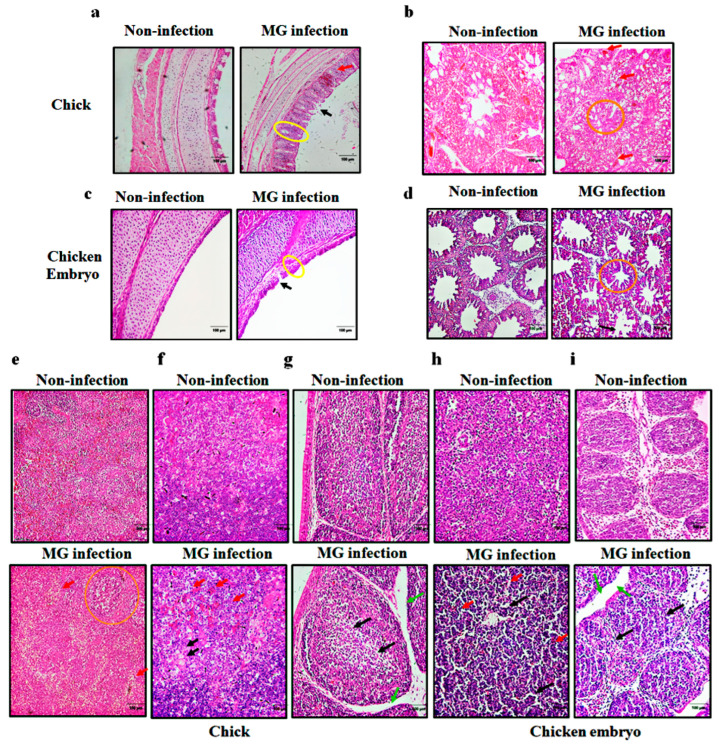
Figure 2.
Effect of MG infection on histopathology of trachea, lung, and immune organs in chicks and chicken embryos. Histopathological changes of trachea and lungs in chicken embryos (a,b) and chicks (c,d). Increased tracheal mucosa thickness (yellow circles); inflammatory cell infiltration (red arrows); an increase in the alveolar space (orange circle); shedding of cilia or epithelial cells (black arrows). (e–i) histopathological changes of spleen and bursa of Fabricius in chicken embryos (e,f) and newly hatched chicks (g–i). Enlarged splenic nodule (orange circle); lymphocyte reduction (black arrows); inflammatory cell infiltration (red arrows); interstitial edema (green arrows). (Magnification: ×20; scale bar: 100 μm; n = 3).

