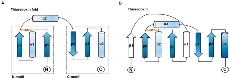
Figure 1.
Schematic structure of the thioredoxin fold and E. coli thioredoxin. (A) The structural elements of the thioredoxin fold are shown in blue. (B) The structure of E. coli thioredoxin is shown with the typical thioredoxin fold in blue and additional features in white. The location of the CysXXCys motif and N-and C-termini are indicated. Arrows represent ß-strands, and α-helices are shown as cylinders (adapted from Martin, 1995 [17]).

