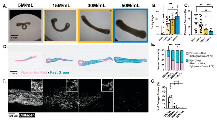
Figure 4.
Structural integrity observed in different meso-ECT dose conditions is correlated with developed prestrain at the tissue level as well as extent and organization of collagen remodeling during tissue formation at the cellular level. (A) Brightfield images of ECTs showing curling in stress-free environment (removed from posts) after 7 days of culture; (B) quantification of ECT curling angle (degrees) and (C) developed prestrain (%); (D) representative histological staining of PRFG for each density condition; (E) quantification of the picrosirius red (collagen content) and fast green (basic proteins, cytoplasm content) per condition measured by % of area analyzed; (F) representative images of SHG imaging showing organized collagen fibrils; (G) quantification of collagen content imaged through SHG for each condition, measured by % area analyzed. 1 mg/mL collagen concentration utilized; M indicates million; n = 7–10 analyzed tissues per group for PRFG staining; n = 12–23 tissues analyzed for curling angle and prestrain quantification per condition; n = 4–7 for SHG with multiple areas averaged to analyze per tissue; the individual points represent single meso-ECTs analyzed; * p < 0.5; ** p < 0.01; *** p < 0.001, **** p < 0.0001.

