Figure 6.
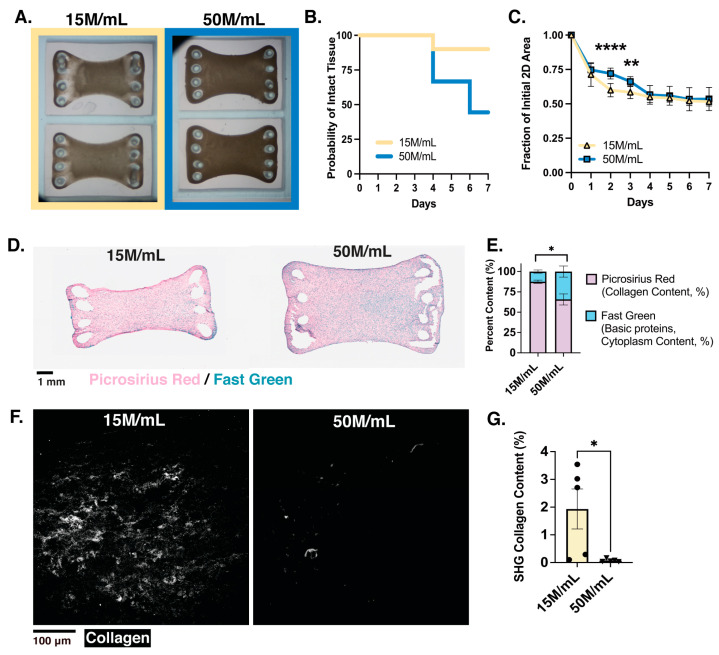
Macro-ECTs maintain dose dependence of tissue survival, collagen content, and organization but have more uniform compaction. (A) Brightfield images show macro-tissue compaction at day 7 (D7) of in vitro culture (mold size, 8 × 12 mm); (B) survival curve of percentage of intact tissues shows structural integrity begins to decline on day 4; (C) quantification of tissue compaction over 7-day culture; (D) representative histological staining of PRFG for each density condition; (E) quantification of the picrosirius red (collagen content) and fast green (basic proteins, cytoplasm content) per condition, measured by % of area analyzed; (F) representative images of SHG imaging showing organized collagen fibrils, measured by % area analyzed; (G) quantification of collagen content imaged through SHG for each condition. Collagen concentration of 3.5 mg/mL utilized; M indicates million; n = 9–10 tissues per condition analyzed for formation; n = 4–6 tissues analyzed histologically with multiple regions averaged per tissue for each condition; the individual points represent single macro-ECTs analyzed; * p < 0.5; ** p < 0.01; **** p < 0.0001.

