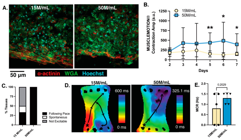
Figure 7.
Increased cell density within macro-tissue format shows limited sarcomere organization but increased contractile amplitude and ability for electrical pacing with no arrhythmia generation. (A) Histological staining of α-sarcomeric actinin (α-actinin), wheat germ agglutin (WGA), and Hoechst; (B) video-based analysis of ECT contractility using the software MUSCLEMOTION® to quantify contraction amplitude; (C) percent of tissues following 0.5 Hz point stimulation pacing during optical mapping; (D) heatmap of activation sequences generated from GCaMP calcium transient recordings for 15 M/mL macro-ECT (left) and 50 M/mL macro-ECT (right); (E) maximum capture rate (MCR) of macro-ECTs under field stimulation. Collagen concentration of 3.5 mg/mL utilized; M indicates million; n = 4–6 tissues analyzed with multiple regions averaged per tissue for each condition for histological analysis; n = 9–10 tissues per condition analyzed for contractility; n = 5–6 tissues per condition attempted for optical mapping, with pacing achieved in n = 2–5; the individual points represent single macro-ECTs analyzed; * p < 0.5; ** p < 0.01.

