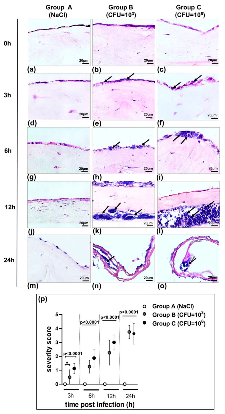
Figure 2.
(a–o) Application of the 3D co-culture model of pleura in studying host–pathogen interactions. The organotypic co-culture model of pleura was infected with S. aureus strain ATCC 49230 for 3 h, 6 h, 12 h, or 24 h with 1 × 10³ and 1 × 10⁶ CFU/mL and tissue damage was analyzed by hematoxylin and eosin staining (a–o). In infection group B we detected bacterial accumulation on the mesothelial cell layer at 6 h post infection (h) and bacteria cells infiltrating soft tissue with formation of mature biofilm under the mesothelial layer (arrows) (k). In infection group C, the accumulation of bacteria on mesothelial cells was more significant than that in group B (i). This was accompanied by a large amount of bacterial cell infiltration and showed numerous, highly active cocci in biofilm formation distributed in the tissue (arrows) (l). Bacterial invasion of the tissue and biofilm building were consistent with tissue damage and capsule formation (arrows) (n,o) after 24 h exposure to S. aureus in both groups without a difference (B and C). Few inflammatory cells infiltrated the pleura in the control group (A). The saline control group (A) was aseptic. Images are from a single experiment and are representative of each group. Scale bar = 20 µm. (p) Histological severity scoring of tissue pathology of the 3D co-culture model of pleura exposed to S. aureus strain ATCC 49230. Histological severity scoring was performed in a double-blinded manner using the following criteria: 0, unaffected tissue; 1, mild injury with minor mesothelial loosening; 2, moderate injury with some mesothelial disruption; 3, severe injury with continuous mesothelial disruption and some detachment; 4, extensive injury, massive mesothelial disruption, and detachment (n = 8). The bars show the mean ± s.d. of two individual experiments. * p = 0.0192.

