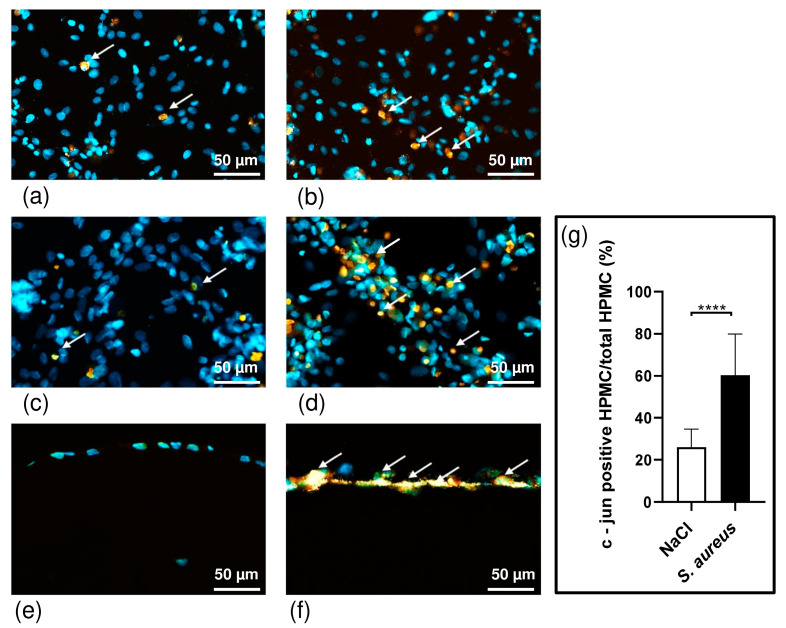
Figure 4.
Immunohistochemically analysis of paraffin-embedded tissue. Immunohistochemistry staining of non-infected HPFs (a), HPMCs (c), and 3D co-culture model of pleura (e) in contrast to infected HPFs (b), HPMCs (d), and 3D model (f). Anti c-Jun (orange) and 4′,6-Diamidino-2-phenylindol (DAPI) (blue). Percentage of c-Jun-positive HPMCs of all cells within the 3D co-culture model. There are significantly more c-Jun-positive cells in the S. aureus infected group (indicated by arrows). The percentage of c-Jun-positive area is significantly higher in the S. aureus group (n = 11) than in the control group (n = 11); **** p ≤ 0.0001, unpaired t-test (g).

