Figure 1.
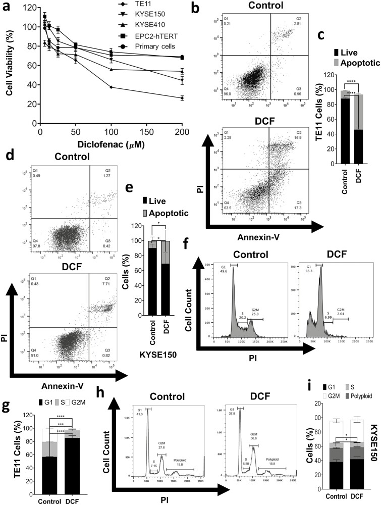
DCF inhibition of cell viability is associated with apoptosis and cycle alterations. (a) Cell viability was measured by MTT assay in ESCC cell lines (TE11, KYSE150, and KYSE410), normal immortalized esophageal keratinocyte cell line EPC2-hTERT, and primary esophageal keratinocytes treated with DCF at indicated doses for 72 h. Dose–response curves are indicated for respective cells. (b–i) TE11 or KYSE150 cells were treated for 48 h with 200 or 400 µM DCF, respectively. Apoptosis was measured by Annexin-V/PI flow cytometry with representative dot plot in (b, d) and bar chart summarizes date from three independent experiments in (c, e). (f–i) Cell cycle was analyzed by PI flow cytometry with representative dot plot in (f, h) and bar chart summarizes date from three independent experiments in (g, i). Data in (c, e, g, i) shown as mean ± SD; *P < 0.05; ***P < 0.001; ****P < 0.0001 by t-test; n = 3.

