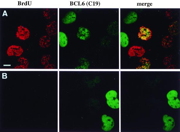
FIG. 5.
Blocking cell cycle progression alters the subnuclear distribution of BCL6. UTA-L cells were synchronized in mitosis and then both induced for BCL6 and exposed to BrdU for 6 h as described for Fig. 3 in the absence (A) or presence (B) of 0.5 mM l-mimosine, which blocks the cells in late G1 (37). Untreated cells expressing BCL6 typically show either nuclear diffuse or nuclear punctate BCL6 staining (green), corresponding to BrdU-negative (presumably in G1) or BrdU-positive (in S; detected in red) cells, as shown in Fig. 4, top or middle row, respectively (A). In contrast, l-mimosine-treated cells are almost completely devoid of BrdU staining, as expected, and most show nuclear diffuse BCL6 staining (B). Bar, 10 μm.

