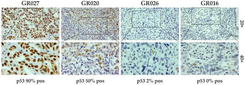
Figure 1.
IHC staining for p53 evaluation. Representative images of samples with different percentages (%) of positive stained cells (brown color; IHC 20× and 40× magnification). In the positive sample, p53 staining was detected in 90% of cells (GR027), whereas the negative ones had percentages of positive cells lower than 70% (GR020, GR026, and GR016).

