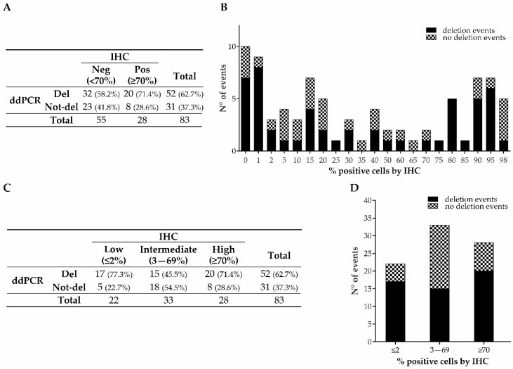
Figure 2.
p53/TP53 status, analyzed by IHC and ddPCR, of retrospective GEAs. Distribution of deleted and not-deleted cases in the groups categorized according to the IHC model based on Gonzalez et al. (A) and in the groups categorized as low, intermediate, and high percentages of IHC p53 staining (C). Distribution of deletion events by ddPCR in the whole cohort (B) and in the three newly categorized groups (≤2% (low), 3–69% (intermediate), and ≥70% (high)) (D).

