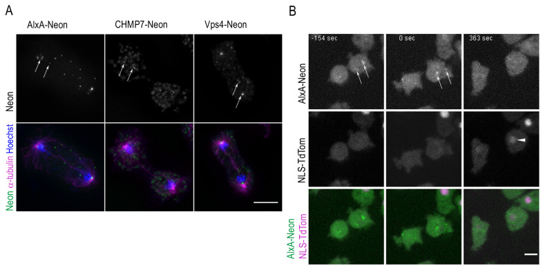
Figure 6.
ESCRT proteins concentrate at the nuclear envelope fenestration sites in mitosis and long after cytokinesis has taken place. (A) Cells were fixed with glutaraldehyde and stained with Hoechst (blue) and anti-α-tubulin (magenta). ESCRT-Neon fusion proteins (green) localize at the nuclear envelope fenestration sites in telophase (arrows). A maximum intensity projection of several slices of the deconvolved Z-stack is presented. (B) Selected time points of Video S13 are shown with cells in cytokinesis (−154 s), after cell division (0 s), and once AlxA-Neon has disappeared together with reappearance of NLS-TdTom (arrowhead) at 363 s. Bars 5 µm.

