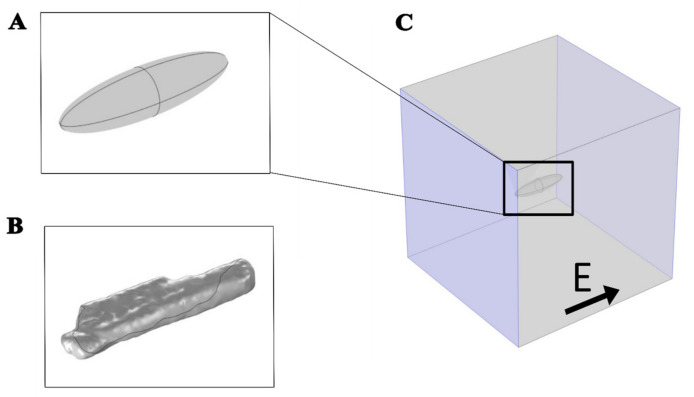
Figure 1.
Geometries used to represent a cardiomyocyte with an electric field applied parallel to the long axis of the cell. (A) Prolate spheroid geometry, 120 µm long, 30 µm wide, and 30 µm high. (B) Real-shaped geometry 142 µm long, 36 µm wide, and 21 µm high. Both the real-shaped geometry and its prolate spheroid approximation were the same as in Milan et al. [30]. (C) The cell was at the center of the box when the electric field was applied parallel to the long axis of the cell. The violet-colored sides of the box represent the electrodes to which the voltage was applied.

