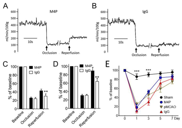
Figure 5.
Functional analysis of M4P in 7-h stroke reperfusion. (A) Exemplary cerebral blood flow from a rat receiving 7-h MCAO. M4P of 100 µg was injected intravenously 1 h prior to recanalization. The blood flow was recorded using a Laser-Doppler flowmetry. (B) Exemplary cerebral blood flow from a rat treated with 100 µg control IgG. (C) Summary of cerebral blood flow from M4P and IgG-treated animals. The blood flow was normalized to baseline. For M4P, n = 9 rats; for IgG, n = 6 rats. (D) Cerebral blood flow from animals receiving 3-h stroke reperfusion. M4P (100 µg) and IgG (100 µg) were injected 1 h prior to recanalization. For M4P, n = 6 rats; for IgG, n = 10 rats. (E) Assessment of motor functions using the Rotarod test (n = 7 rats/group). Statistical analysis was performed by two-way ANOVA with Bonferroni’s post hoc analysis. ** p < 0.01, *** p < 0.001. The numerical data supporting the graphs can be found in Supplementary Tables S6–S8.

