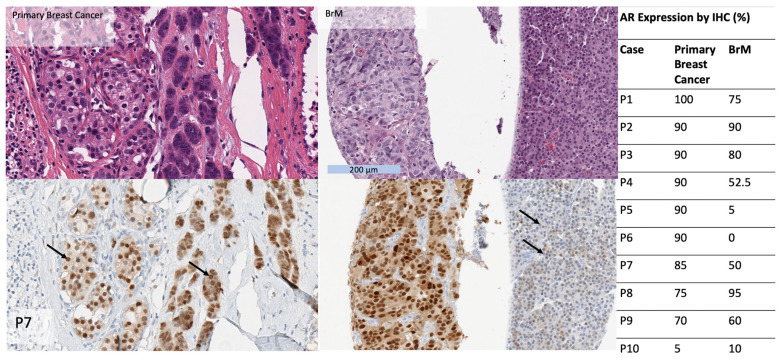
Figure 3.
Table (right): AR expression by IHC (in %) in BrM compared to matched primary breast tumours, in a subset of 10 patients. Images show an example of a primary breast cancer and BrM from the same patient, hematoxylin and eosin (top) and SP107 IHC (bottom). The primary and BrM show morphologic heterogeneity with cells with lower nuclear grade and relatively abundant cytoplasm and cells with higher nuclear grade and scant cytoplasm. AR expression is diffusely positive in both components in the primary tumor but AR expression is diminished in the higher grade component in the BrM (arrows).

