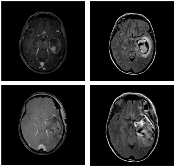
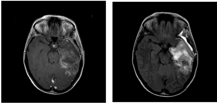
Figure 1.
Example MRI images of a patient with temporal glioblastoma: (top) 2 days before surgery, (middle) 2 days after surgery, and (bottom) 17 days after surgery (for radiation treatment planning). T1-weighted contrast-enhanced (Gadolinium) images on the left and T2-weighted FLAIR images on the right. MRI = magnetic resonance imaging; FLAIR = fluid-attenuated inversion recovery.


