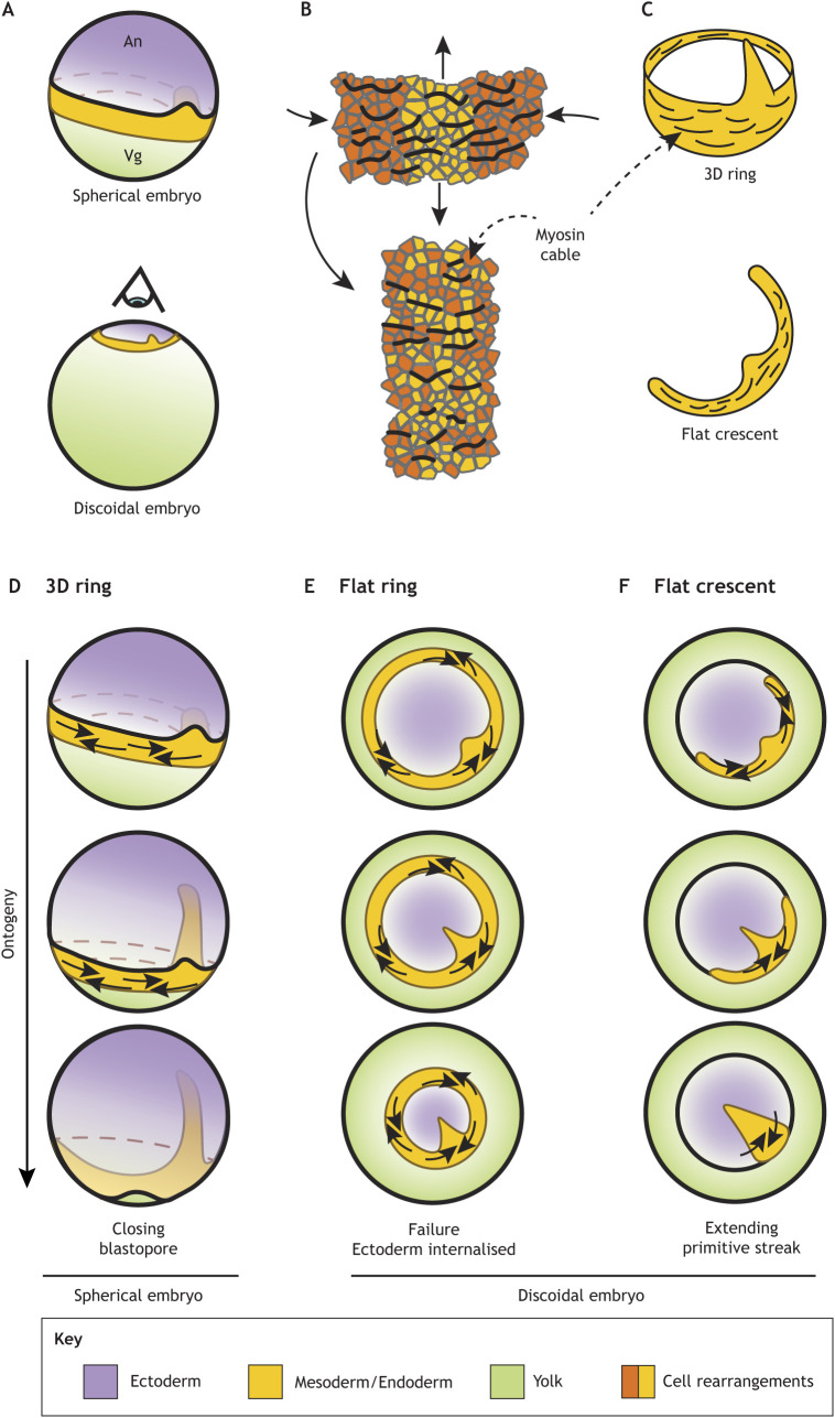
Fig. 4.
Mesoderm-ring to -crescent transition. (A) The increase in yolk size resulted in the flattening of the embryos during amniote evolution. The eye indicates the direction of the projection in E,F. (B) The direction of actomyosin cables is perpendicular to the direction of intercalation. Directed cell intercalation drives the convergent extension of tissue. (C) Actomyosin cables are organised along the long axis of the prospective mesoderm in tetrapods. (D-F) Mesoderm undergoes convergent extension in three different scenarios. In a 3D ring, the contraction of the ring does not interfere with the ectoderm expansion (D). A flat mesoderm ring undergoing convergent extension traps the ectoderm, which prevents its expansion (E). A flat mesoderm crescent undergoing convergent extension naturally collapses into the midline (F).

