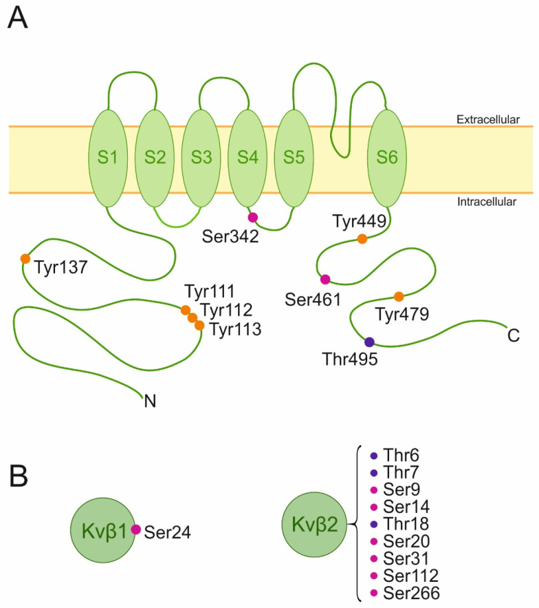
Figure 1.
Schematic representation of Kv1.3 membrane topology and Kv1.3 beta ancillary subunits highlighting putative phosphorylation residues. (A) The channel presents six transmembrane domains (S1–S6) and intracellular N and C terminal domains. (B) Cytosolic Kvβ subunits. Phosphorylated amino acids are highlighted with the three-letter code and colored dots. Magenta (serine), purple (threonine), and orange (tyrosine) [141].

