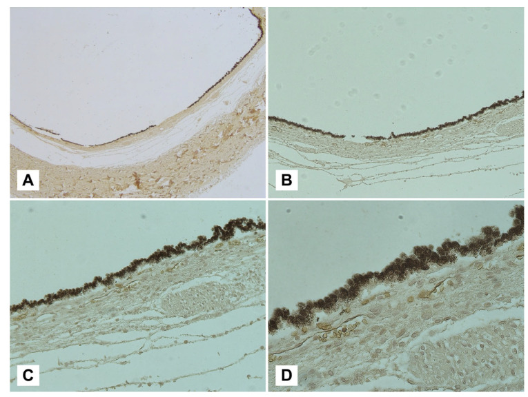
Figure 4.
Visualization of a sub-retinal plane with glycophorin-A immunolabeling at different magnifications: (A) 2.5×; (B) 5×; (C) 10×; (D) 40×. Following the removal of the retinal membrane, a strong immunopositivity for the membrane antigen glycophorin-A can be observed at the level of the choroidal membrane, interpretable as sub-retinal blood collection.

