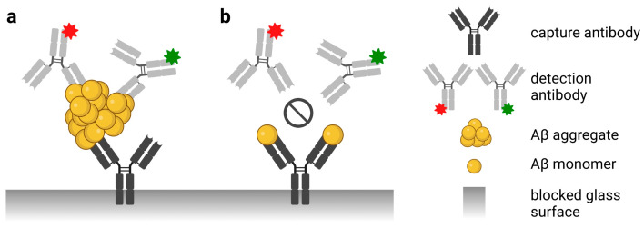
Figure 1.
Scheme of the sFIDA principle. The biochemical principle of sFIDA is similar to a sandwich ELISA with capture and detection antibodies directed against the same or overlapping epitopes of the N-terminus of Aβ. Monomeric and oligomeric Aβ species of the sample bind to the capture antibodies. (a) However, the red or green fluorescently labeled detection antibodies only detect aggregated Aβ species such as oligomers because the assay antibodies bind to the same or overlapping epitope. (b) Therefore, the red or green labeled detection antibody cannot bind monomers because the capture antibody already masks the epitope. Subsequently, the assay surface is imaged using dual-color fluorescence microscopy (excitation at 635 and 488 nm), and only colocalized pixels above a defined cutoff threshold are counted by image data analysis. Abbreviations: Aβ, amyloid-β; sFIDA, surface-based fluorescence intensity distribution analysis. Created with BioRender.com (accessed on 26 April 2023).

