Figure 3.
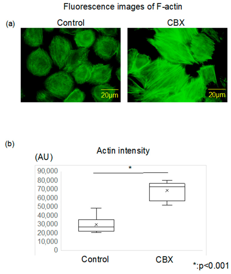
Actin polymerization and intensity between control and CBX treatment. (a) Actin cytoskeleton was stained with Alexa 488-conjugated phalloidin. The actin filaments in the control group were shorter, less organized, and oriented randomly. Conversely, in comparison with the control cells, the actin filaments in cells of the CBX treatment group were distributed throughout the cell and aligned along the long axis of the cells. (b) Actin staining intensity was evaluated via arbitrary units (AU) using Image J software. Actin intensity in the CBX treatment group was significantly higher than that of the control group.

