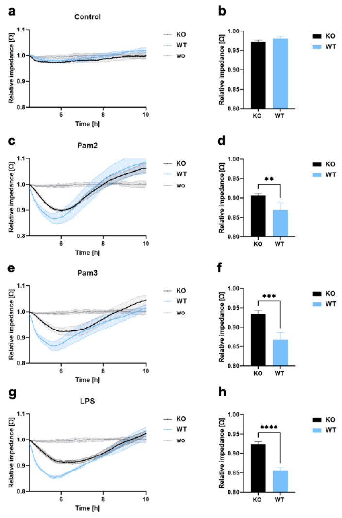
Figure 2.
Endothelial barrier disruption and transmigration in THP-1 WT and KO cells. (a–h) LECs were seeded onto ECIS arrays (96W20idf PET) to form an endothelial monolayer, and 1 × 10⁵ THP-1 KO or WT cells were added (a,b), either without a ligand or with (c,d) Pam2, (e,f) Pam3, or (g,h) LPS. Monolayer disruption was documented in real time by impedance measurements using the ECIS system (9600Z). Time course diagram and bar graphs at 6 h after treatment showing mean values ± standard deviation (n = 4). A t-test was performed to assess differences between KO and WT cells (** p < 0.01; *** p < 0.001; **** p < 0.0001).

